 Assisted reproductive techniques (ART), or assisted reproductive techniques (ART) in English, are procedures based on the logic of taking the egg cells produced in the female body out of the body with special needles, fertilizing them with the man's sperm in a laboratory environment, and transferring the resulting embryo or embryos into the female uterus.
With the use of assisted reproductive techniques, many couples can have babies today. Assisted reproductive techniques in modern medicine are classical in vitro fertilization (IVF) and microinjection, also known as intracytoplasmic sperm injection (ICSI). Assisted reproductive techniques in modern medicine classic in vitro fertilization or in-vitro fertilization (IVF) and microinjection or otherwise known asintracytoplasmic sperm injection (ICSI) .
The only difference between in vitro fertilization and microinjection is the mode of fertilization.
Microinjection, or ICSI for short, is one of the latest advancements in assisted reproductive techniques. With this method, sperm directly enters the egg. With the introduction of ICSI, the chances of treating in vitro fertilization and especially infertility due to male problems have increased considerably and new horizons have opened.
TESE AND TESA The methods called are techniques used for cases where there is no sperm in the semen sample or when sperm is produced but cannot be expelled. For example, in cases where the man's ducts are blocked and he cannot ejaculate the large amount of sperm in his testicle, tissue is taken from the man's testicle with a needle, sperm are found in it and fertilization is achieved with the sperm obtained. This procedure is called testicular sperm aspiration, TESA for short. Or, a piece/tissue sample is taken directly from the testicle and sperm is obtained from this tissue, which is called testicular sperm extraction (TESE).
While the TESE procedure used to involve roughly removing one or two pieces of tissue from the testicle, now the procedure is performed using an operating microscope. In this process microTESEIt is said. Compared to the classical TESE application, the chance of finding sperm is higher and the possibility of damaging the testicle is lower. In testicular sperm aspiration (TESA), sperm is obtained by aspirating, that is, by pulling it with negative pressure, in patients with blocked ducts. In all in vitro fertilization applications, the common goal of the different ovulation treatments used is to produce a large number of eggs. This treatment includes controlled ovarian stimulation.
In Which Situations Are In Vitro Fertilization Applications Performed?  • In women with blocked uterine canals (tubes)
• In cases where sperm functions are severely impaired
• Women who have widespread adhesions in the abdomen due to endometriosis and cannot achieve pregnancy with treatment.
• Immunological (concerning the immune system) Infertility
• In Some Hormonal Disorders
• In cases where pregnancy cannot be achieved with other treatment methods
• Infertility whose cause cannot be explained by the tests performed
• In order to obtain a healthy baby by diagnosing some hereditary diseases at the embryo stage (together with pre-term genetic diagnosis methods).
• In order to select healthy embryos by genetic diagnosis method in women with recurrent miscarriages.
• If pregnancy cannot be achieved despite the insemination method being applied several times.
WHAT ARE THE STAGES OF IVF?
Egg Development
Various stimulating hormone drugs are used to ensure egg development. In classical practice, during the preparation period to control egg development with externally administered hormonal drugs, approximately for two weeks suppressive hormones are used. These hormones can be used as a subcutaneous injection or nasal spray that the person can apply himself. Following suppressive treatment, the patient who menstruates within 10-14 days is evaluated with blood tests and ultrasonography, and hormonal drugs that will stimulate egg development are started. These suppressive injections are given to the person in order to prevent defects that may occur in the nervous system of the fetus when pregnancy occurs. folic acid support is also provided. In addition, both partners are given short-term antibiotic treatment in order to eliminate some possible microbes that may negatively affect the treatment. Purpose of stimulating needles used in assisted reproductive techniques lots of eggs and then lots of embryos to increase the chances of pregnancy. A separate treatment and dosage schedule is determined for each patient, depending on the woman's age, ovarian reserve, blood hormone values and height/weight ratio.
Examples of applied protocols:
SHORT PROTOCOL GnRHa application begins on the first day of menstrual bleeding and continues until the end of the treatment (the day of the cracking shot).
ULTRA SHORT PROTOCOL GnRHa is started on the first day of menstrual bleeding and stopped after three days. Treatment is continued with hMG or FSH.
ANTAGONIST PROTOCOL Stimulant drugs are started within the first few days of menstrual bleeding, and a few days later, treatment is continued by adding drugs called antagonists that prevent premature cracking.
LONG PROTOCOL GnRHa administration begins approximately on the twenty-first day of the previous period. On the third day of the following menstrual bleeding, it is determined by a blood test whether there is suppression or not. If the blood estrogen level has decreased, it means that suppression has been achieved. In this case, stimulation therapy is started, but GnRHa administration is not terminated. It continues until the day the cracking shot is given.
Which protocol will be suitable for you and what dose of medication will be used is decided at the first examination. Following the initiation of stimulant medications ultrasonography and blood testsThe expectant mother is followed up. Although it is less common in the beginning, it sometimes occurs after egg development reaches a certain level. daily checks must. Appropriate changes are made in drug doses in line with the follow-ups. Egg development takes 10-12 days on average. When the eggs reach a certain size, cracking needle The last injection, called the so-called egg, is applied and approximately 36 hours later, the eggs are taken out of the body with the help of special needles before they hatch and are delivered to the laboratory. It should be known that in in vitro fertilization pregnancies obtained with the use of these drugs, the development of a disabled baby, which is thought to be due to the drugs, has not been observed.
Does each follicle in the ovaries contain an egg?
No. However, if there are many follicles, it becomes difficult to count all of them. In this case, more eggs can be obtained than what we see on ultrasonography. However, unfortunately, in cases where only one or two follicles are present on ultrasonography, eggs may not be obtained. The average fertilization rate is 70-80%. While some couples have a higher rate of fertilization, unfortunately there is rarely any fertilization at all. In this case, the couple is informed about the situation and the transaction is cancelled.
Egg Collection Process
Egg collection (OPU - Oocyte pick up) is a procedure performed vaginally with the help of a needle added to the Ultrasonography (USG) probe, at the 36th hour following the application of the egg cracking needle. It is decided when the eggs will be collected by looking at the level of estradiol hormone in the blood and follicle diameters. Depending on the patient's wishes, local or general anesthesia can be applied. If general anesthesia is requested, the patient must be informed in advance as he/she will not have to eat or drink anything since the previous evening. The ovaries are reached with the help of a needle under vaginal USG, and each follicle is aspirated one by one and taken into sterile tubes. The tubes are given to the embryology laboratory and the presence of eggs is investigated. If no egg is found in the first aspiration from the follicle, the follicle is washed. After the egg is found, it moves on to other follicles. The egg collection process takes approximately 15-20 minutes and is a procedure that can be tolerated by patients and does not cause serious pain. Since groin and abdominal pain may sometimes occur, there is no harm in taking paracetamol-type painkillers. One of the situations that may be encountered during the procedure is small bleeding that may occur where the needle enters, and the other is infection. Bleeding is eliminated by simple tamponade. Infection is controlled with antibiotic treatment. However, this rate is less than 1%.
The follicle fluid given to the embryology laboratory in a tube is evaluated under a microscope by the embryologist and the doctor is informed. The egg seen in the follicular fluid is removed with the help of a pipette, washed in special media and placed in another petri dish. Before starting the microinjection process, it is kept in the incubator for 2 hours.
When deciding whether in vitro fertilization (IVF) or microinjection will be applied to the collected eggs, the patient's age, egg number, quality, sperm status (number, movement and shape), whether there have been previous attempts, and whether PGD will be applied or not are evaluated. Even if the decision to apply in vitro fertilization is made, some of the eggs are usually microinjected. This is to prevent an unexpected fertilization failure.
In Vitro Fertilization (IVF) If the application is decided, 4 hours after the eggs are collected, the eggs and washed sperm are compared in drops in a tube or in a culture petri dish. This process is done by calculating the number of sperm per egg. In this process, the sperm carries out fertilization itself. Fertilization control is performed after 18-20 hours.
When sperm parameters are below normal limits, e.g. In cases where the number is low, motility is low, or the normal morphology is below 4%, the sperm's ability to fertilize eggs is very low. Even in more advanced cases, which we call severe male infertility, the sperm cannot fertilize the egg itself. In this case, Microinjection should be preferred. Microinjection is also suitable in cases where the number of eggs is low or egg quality is low. Microinjection is even preferred in patients whose sperm and egg quality is good but a low fertilization rate has been observed in previous applications.
The eggs to be microinjected are first subjected to mechanical and enzymatic processes and cleaned from the surrounding cells called cumulus. The maturity of the cleaned eggs is evaluated. The egg called Metaphase II is the mature egg that we will apply microinjection to. Since the metaphase-I egg is not yet fully mature, microinjection cannot be performed. Prophase-I egg is an immature egg with a germinal vesicle. Therefore, microinjection cannot be performed. If Metaphase-I eggs develop during the day and become Metaphase-II, microinjection is performed. The eggs are also evaluated by the embryologist from a cytoplasmic perspective.

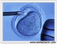
Microinjection is performed under a microscope, which we call a micromanipulator. 2 glass pipettes prepared at certain angles are used. One of these is the holder pipette that fixes the egg, and the other is the injection pipette that we inject into the egg after capturing the sperm and immobilizing it. Here, the fertilization rate is higher than the IVF method since the sperm is released directly into the egg.
After the process is completed, the eggs are transferred to a new medium and placed in the incubator. Fertilization control is performed after 18-20 hours. When checking fertilization, it is expected that there will be 2 pronuclei (nuclei), one coming from the egg and the other coming from the sperm. Each egg is evaluated individually and any features seen are noted. After the eggs are cleaned, each evaluated oocyte is followed step by step until it is transferred.

The natural nutritional needs of the embryo and the source from which it meets this need change over time. To achieve this in the environment outside the body, different media are used in different developmental periods.
Embryo Transfer:
In embryos where fertilization is checked, it is checked whether there is division on the 2nd day. If division has occurred, embryo quality is evaluated by looking at how many cells (blastomeres) it has, the equality of blastomeres, fragmentation rate and some other parameters. Embryo transfer can be done any day from the 2nd to the 5th day. The date of transfer is decided by taking into consideration the embryo quality, the number of developing embryos, the age of the patient, whether genetic diagnosis is made or not. At our center, transfer is usually made on the 3rd day. However, in cases where the embryo quality is good and the number of embryos is high, and the patient's age is appropriate, we prefer to transfer the embryo at a later stage, namely the blastocyst stage, on the 5th day in order to prevent multiple pregnancy. During the transfer process, the patient is laid in the examination position and then the secretions called mucus in the vagina and cervix are cleaned with sterile culture fluids. The best embryos are selected and placed in the mother's uterus with the help of a transfer catheter under ultrasound guidance. This procedure is a short-term, painless procedure. Information is given about the condition of the embryos, and after a short rest, medication information is given and the patient is sent home.

Assisted Hatching:
There is a shell-like membrane structure around the embryo that protects it, which we call zona pellucida. During natural development, when the embryo reaches a certain size, this membrane opens (hatching) and the embryo attaches to the inner wall of the uterus. This membrane can sometimes be thick. In this case, it becomes difficult for the embryo to attach to the inner wall of the uterus. To reduce this risk, the zona pellucida is thinned in a certain area. This process is called assisted hatching. Mechanical, chemical or laser methods can be used. In our center, laser method is used for thinning process.
Embryo Freezing:
If there are many high-quality embryos belonging to the patient other than the transferred embryos, they can be frozen. By thawing and transferring frozen embryos in later applications, the woman's need for retreatment is eliminated. At this stage, the patient is given only drugs that prepare the inside of the uterus. This provides both psychological and economic advantages. Embryos that are decided to be frozen are gradually balanced with chemicals called cryoprotectants to prevent damage during the freezing and thawing processes. Two methods can be applied to freeze embryos.
1)Slow freezing
2)Fast freezing (vitrification)
After the embryos are frozen, they are stored in liquid nitrogen at -196 C until thawed. Since ice crystals that may form due to the liquid content of the embryo while cooling the embryos to a temperature of -196 degrees Celsius may damage the integrity of the embryo, the cooling process is carried out gradually by using protective chemicals in the slow freezing technique. In vitrification, a vitreous or glassy structure is created in which ice crystals do not form at all, and the cells are frozen by dipping them directly into liquid nitrogen. When thawing frozen embryos, the opposite process is applied, using special protective chemicals. If it is observed that the thawed embryos continue their vitality and grow, they are transferred. Embryos may be damaged during the freezing and thawing processes, not during the time they are kept in liquid nitrogen.