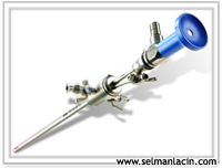
 hysteroscopy It is the monitoring of the cervix and uterine cavity by placing an optical device into the uterus through the vagina. The tool or tools used during this process hysteroscope It is called.
Such as investigating the causes of infertility, investigating the causes of abnormal vaginal bleeding, and diagnosing congenital abnormalities. for diagnostic purposes or removal of previously detected myomas or polyps, surgery for congenital deformities, etc. for therapeutic purposes Hysteroscopy can also be performed.
Procedures in which hysteroscopy is most commonly used removal of the septum within the uterus, opening of adhesions, removal of polyps, evaluation of in vitro fertilization failure and myoma removal operations.
formed in the uterus adhesions It is one of the important reasons for not being able to get pregnant, miscarriage and having little or no menstrual period. It is also called Asherman syndrome, after the person who first described it. It usually follows a damage caused in the uterus. The most common reason is the decrease in the amount of menstrual bleeding after an abortion or the lack of menstruation, which is understood to be an adhesion in the uterus in research. It is especially likely to occur after "too energetic" abortions. Other reasons following abortion include interventions due to the baby's partner not coming out completely after birth and surgeries for fibroids in the uterus.
Less commonly, it can also be seen after childhood tuberculosis (TB) or another infection. As a result of damage to the cell layer that is responsible for renewing the inner layer of the uterus every month after abortion, the inner layer of the uterus can no longer be renewed and the walls of the uterus stick together.
What are the symptoms and how is the diagnosis made?
Most commonly, menstrual bleeding stops or decreases significantly after abortion. Normally, for women who have regular menstrual periods after an abortion, menstrual bleeding is expected to occur within 4-8 weeks. If it does not come, uterine adhesions should be suspected. Less commonly, painful menstrual bleeding, inability to become pregnant without any reason, and recurrent miscarriages may also be symptoms of intrauterine adhesions. Vaginal ultrasound examination, uterine film (HSG), and hysteroscopy are tools that can be used for diagnosis. Among these, hysteroscopy, which confirms the diagnosis and provides treatment in the same session, is considered the gold standard.
How is it treated?
The current standard in the treatment of intrauterine adhesions is hysteroscopic adhesion removal. Abortions that were performed in the past are now history. Intrauterine adhesions can be opened using scissors or electrical energy. The success of the procedure is directly proportional to the degree of adhesions and the experience of the surgeon. The denser the adhesions and the proportionally larger part of the uterine cavity they cover, the higher the chance of treatment. It is sometimes not possible to treat adhesions, especially those that completely cover the uterine cavity. In cases where adhesions are intense, sometimes more than one attempt may be required. After hysteroscopy, a balloon or spiral can be placed in the uterus to prevent adhesion from forming again and taking up space. After the procedure, hormone therapy is also given for cell renewal. In those with severe adhesions, it would be useful to have a hysteroscopy or at least a uterine film taken after menstrual bleeding after the end of hormone therapy.
What is the success of the treatment?
More than one hysteroscopy is often required in women with severe adhesions. In women whose uterine cavity is at an acceptable size or fully dilated, there will be a chance of spontaneous pregnancy if the tube exits are also open. If the uterine cavity has been clearly formed but the tube exits cannot be seen, in vitro fertilization should be performed.
Implementation of the process
If the patient will undergo hysteroscopy for diagnostic purposes only, operating room conditions and general anesthesia may not be necessary. For the procedure, the patient lies on the gynecological examination table and local anesthesia is applied after the vagina and cervix are cleaned with antiseptic liquids. Then, a 0.5 cm diameter incision is usually made without the need for dilation of the cervix. optical device It is slowly placed inside the uterus and at the same time carbon dioxide gas or liquid is injected and the inside of the uterus is monitored on the monitor. Tissue samples are also taken from abnormal tissues for diagnosis. to pathological examination is sent. Patients generally do not feel enough pain to interrupt the procedure and hysteroscopy is completed within 5-10 minutes.
Operative (surgical) hysteroscopy It is performed under similar conditions as laparoscopy. Since the optical instrument and device used here are thicker, the appropriate surgical procedure is performed after the cervix is widened and placed inside. Depending on the nature of the procedure, it may take between 15 minutes and 1 hour.
Are There Risks of Hysteroscopy?
Diagnostic hysteroscopy has very few complications and almost never causes a life-threatening problem. Since operative hysteroscopies are more difficult procedures that require a little more experience, complications occur at a rate of 1 - 2/100. Uterine perforation is the most common complication during the procedure, but the holes usually heal on their own with monitoring and drug treatments, without the need for any additional surgical intervention. When operative hysteroscopy is performed, it is often done to simultaneously see the external structure of the uterus and to prevent possible complications. laparoscopy can also be applied. This issue is a decision to be made by the physician based on the difficulty of the procedure to be performed and his/her experience.
Some other complications may also occur due to the fluids used to expand the uterus during the procedure. These include serious allergic reactions, decrease in body temperature, clotting problems, breathing difficulties, and fluid accumulation in the lungs (pulmonary edema). However, such a problem is rarely seen if the procedure time is not too long, the amount of fluid taken is strictly controlled by the surgeon and with experience.
After Hysteroscopy.
It is normal to experience some vaginal discharge and cramping in the few days following hysteroscopy. Simple painkillers can be used for this. Since the cervix is widened to perform hysteroscopy, sexual intercourse should be avoided for a few days to prevent infection. Normal activities can usually be resumed within a day.
In our center hysteroscopy The process is frequently applied for different purposes.