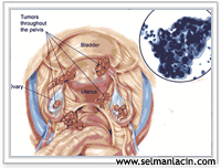
 The most feared type of cancer among women is ovarian cancer. The reason for this is that, since the ovaries are intra-abdominal organs, they do not cause any noticeable findings in their early stages and are usually noticed during lower abdominal examination when they enlarge.
According to statistics, ovarian cancer occurs in one in 70 women. If not diagnosed early, ovarian cancer eventually becomes painful and large, and the fluid secreted by the tumor can cause the abdomen to swell (this is called ascites). In 70–80 percent of cases, the disease is diagnosed at this advanced stage.
Symptoms: There are usually no symptoms at first; Mild discomfort in the abdomen and mild indigestion; swollen abdomen and pain below the abdomen (in the advanced stage of the disease). Ovarian cancer usually develops after menopause. However, it can rarely be seen at young ages. Women who have never been pregnant or have had problems getting pregnant are more likely to get ovarian cancer. Conversely, this is less likely if you have had many children or have used birth control pills.
Conditions thought to reduce the risk of ovarian cancer: Birth control medication use, childbearing and breastfeeding, removal of the uterus, ligation of the tubes, removal of the ovaries as a preventive measure, avoidance of fatty diets, but in all cases, annual lower abdominal examination is very important. Because early diagnosis and treatment provides a much greater chance of recovery.
Diagnosis If a mass in the ovaries is noticed during the lower abdominal examination and gynecological examination, the doctor orders some tests. Ultrasound may be required to understand the condition of the inside of the abdomen, x-rays of the large intestine, kidneys and ureters (the channels connecting the kidneys to the bladder), and rarely laparoscopy (which is performed by the doctor making an incision in the abdomen and inserting an instrument inside) to see the condition of the ovaries may be required. The doctor may take a small piece of the tumor (the only way to tell if it is malignant) during laparoscopy. Three-quarters of ovarian tumors are nonmalignant.
Women who have relatives with ovarian cancer and who are at high risk of developing ovarian cancer can be screened with transvaginal ultrasonography (an ultrasound method performed with a small instrument inserted into the vagina) and blood tests. Transvaginal ultrasonography displays mass formations in the ovaries without determining whether they are benign or malignant. Some features of ovarian cysts may suggest that these cysts may be more malignant tumors.
Blood tests for ovarian cancer include: CA–125 There is a level measurement. The blood level of this protein is increased in women with ovarian cancer. However, it should not be forgotten that CA-125 may also increase in the blood in some non-cancerous ovarian diseases such as infection and endometriosis. Additionally, CA-125 may not increase in some rarer ovarian cancers. When this test is found to be high, further examinations (imaging methods) should be performed, and if fluid (ascites fluid) is detected in the abdominal cavity, it should be investigated whether there is cancer by taking a sample of this fluid and examining it or by taking a sample directly from the ovaries. When necessary, the ovaries are monitored using methods such as computerized tomography or MRI (magnetic resonance).
Computed Tomography (CT): It is an imaging method in which the X-ray rotates around the human body and images from various angles. The images taken are then combined by a computer to create a detailed image of the inside of our body. As the film is shot, details are displayed in greater detail using a contrast agent, which can be thought of as a type of dye.
Magnetic resonance imaging (MRI): It gives detailed pictures of the inside of our body using a magnetic field and a computer. X-rays are not used in this method.
Ultrasonography: It is performed by reflecting the images obtained by the sound waves released from a device called a probe, from the ovaries or other organs, onto the screen. It distinguishes whether the structure of mass formations is solid or fluid-filled cystic structure. It is the most commonly used imaging method.
Color doppler current: This method shows the blood supply of the ovaries through vessels. In ovarian cancers, blood flow to the ovaries generally increases and Doppler measurements are found to be increased. However, in some benign diseases, blood flow in the ovaries may increase. In women, blood CA-125 testing and transvaginal ultrasonography are not fully sufficient for the definitive diagnosis of ovarian cancer. For definitive diagnosis pathological examination It is necessary.
Tumor types and behavior of the tumor Cancers are named according to the type of cells in which they develop, show different behavioral characteristics according to the characteristics of these cells, and their treatments may be different. From this perspective, there are cells with very different characteristics in the ovaries. There are approximately ten types of ovarian cancer. The most common are tumors arising from the tissue covering the surface of the ovary. After these come tumors that develop from sex cells. Generally, ovarian cancers spread superficially. Cancer cells shed from the cancer tissue that develops from the ovaries in the abdomen spread within the abdomen by clinging to the surfaces of other organs in the abdomen. It can spread to the tubes, other ovaries, the surface of the intestine and peritoneum, the omentum tissue, the surface of the liver and spleen, and under the diaphragm. The spread of ovarian cancer to distant organs through the blood occurs in very advanced stages.
treatment
Surgery: Surgical Treatment of Ovarian Cancer The abdomen should be opened through a midline incision and a sample of the abdominal wash fluid or ascitic fluid, if any, is taken. The mass must be removed and evaluated pathologically using the frozen section method. If malignancy is detected, surgery should be continued, taking into account the patient's age and whether he or she has children. The aim is to perform the surgical procedure without leaving any tumor behind in the abdomen. The remaining tumor tissue is the factor that most negatively affects the prognosis of the disease in cases of ovarian cancer. If the tumor is limited to a single ovary and the patient's fertility is desired to be maintained, in addition to removing the ovary on the side where the mass is located, while preserving the uterus and the other ovary, omentectomy (abdominal membrane removal), removal of the lymph nodes in the abdomen (lymphadenectomy) and multiple biopsies should be performed from suspicious areas in the abdomen, if any, or from random peritoneal surfaces. After fertility-sparing surgical staging, the surgical procedure performed in stage 1a and grade 1-2 cases is sufficient and there is no need for postoperative chemotherapy. In cases where fertility preservation is not desired, hysterectomy (uterus removal), bilateral ovarian removal, omentectomy and lymphadenectomy should be performed.
Chemotherapy (drug therapy)
The physician applies treatment using a combination of many medications (usually triple combined therapy). After chemotherapy, the remaining tumor tissue usually shrinks. However, after 12–18 months, if complete treatment has not been achieved, the disease may begin to grow again as it develops resistance to drugs. In this case, a different drug combination is used in the second chemotherapy application.
Radiation: Surgery is usually followed by chemotherapy. If the doctor cannot remove the entire tumor, radiotherapy shrinks it, giving chemotherapy a better chance.
Survival: If ovarian cancer is diagnosed early, the chance of survival for 5 years is 60-80 percent. Since this type of cancer cannot be diagnosed at an early stage, the complete recovery rate is 30-40 percent. Despite everything, women can live much longer today, even in advanced cases, with extensive surgery and chemotherapy.